| DREAM OCT VG200D | ||
|---|---|---|
| OCT Imaging | ||
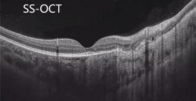
| Methodology | Swept-Source OCT | |
| OCT Central Wavelength | 1030~1070 nm | |
| Scan Speed | 200 kHz (200,000 Hz) | |
| Axial Resolution (Optical) | 5.5 μm | |
| Lateral Resolution (Optical) | 15 μm | |
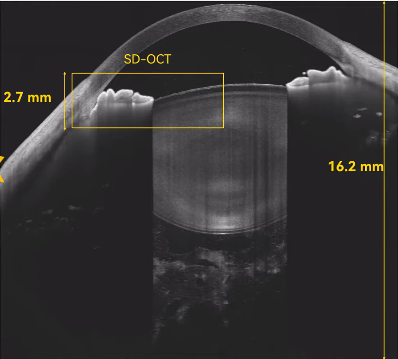
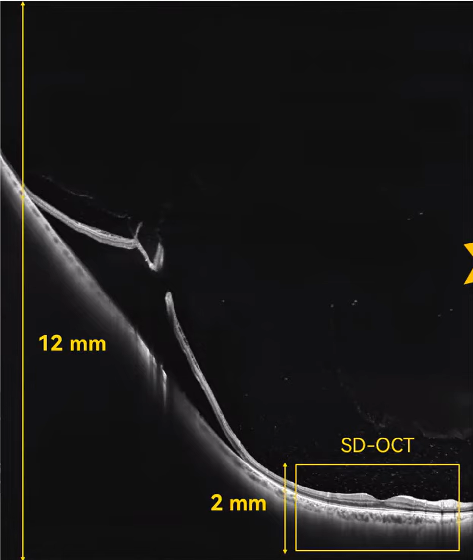
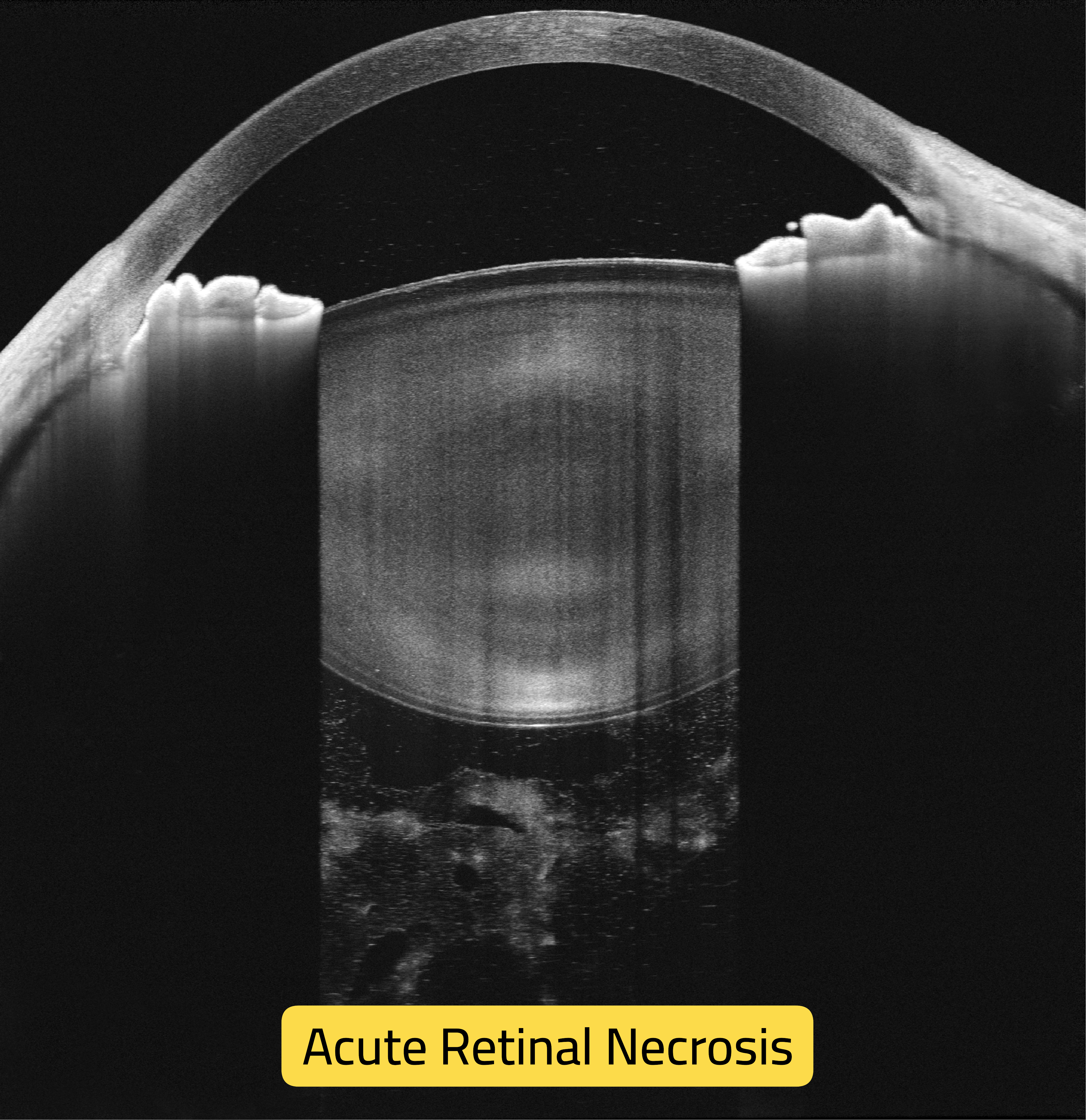
| A-Scan Depth | 1. Retina: 12 mm 2. Anterior segment: 16.2 mm |
|
| Scan Range (Retina) | Without widefield lens: 83° (16 mm) With widefield lens: 130° (26 mm) | |
| Scan Range (Anterior) | 20 mm | |
| OCTA Imaging | ||
| Scan Range (Retina) | Without widefield lens: 83° (16 mm) With widefield lens: 130° (26 mm) | |
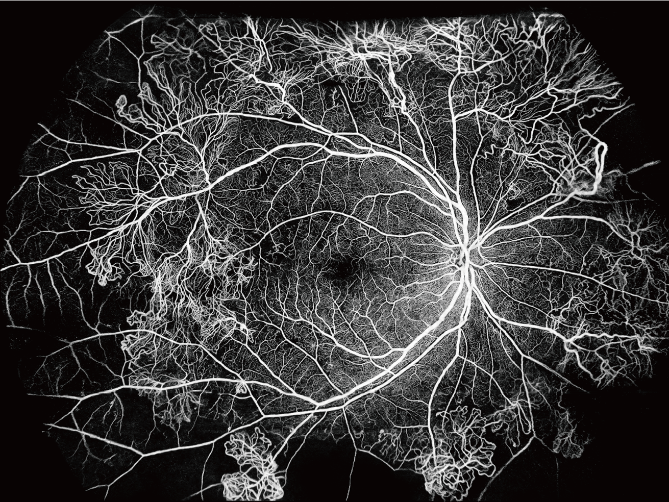
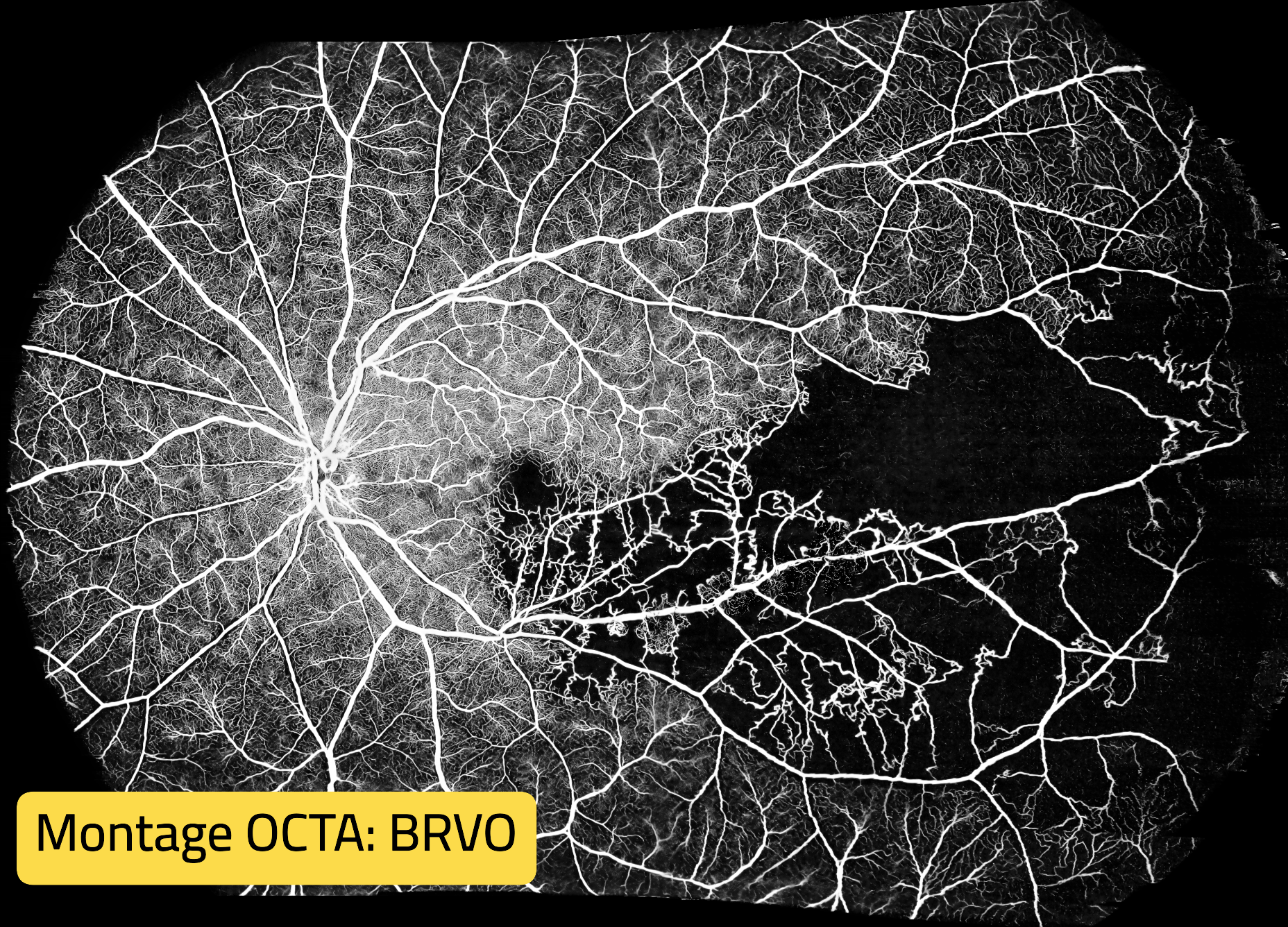
| OCTA Montage | 225° (44 mm × 42 mm) | |
| Algorithm | TRUE Angio™ | |
| Fundus Imaging | ||
| Methodology | cSLO | |
| Optical Source | SLD | |
| Wavelength | 830±20 nm | |
| Field of View | 90° × 90° | |
| Others | ||
| Range of Refractive Compensation | -33 D ~ +40 D | |
| Alignment | Automatic / Electrical | |
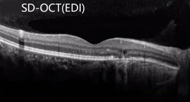
| Feature | Swept-Source OCT | Spectral-Domain OCT |
|---|---|---|
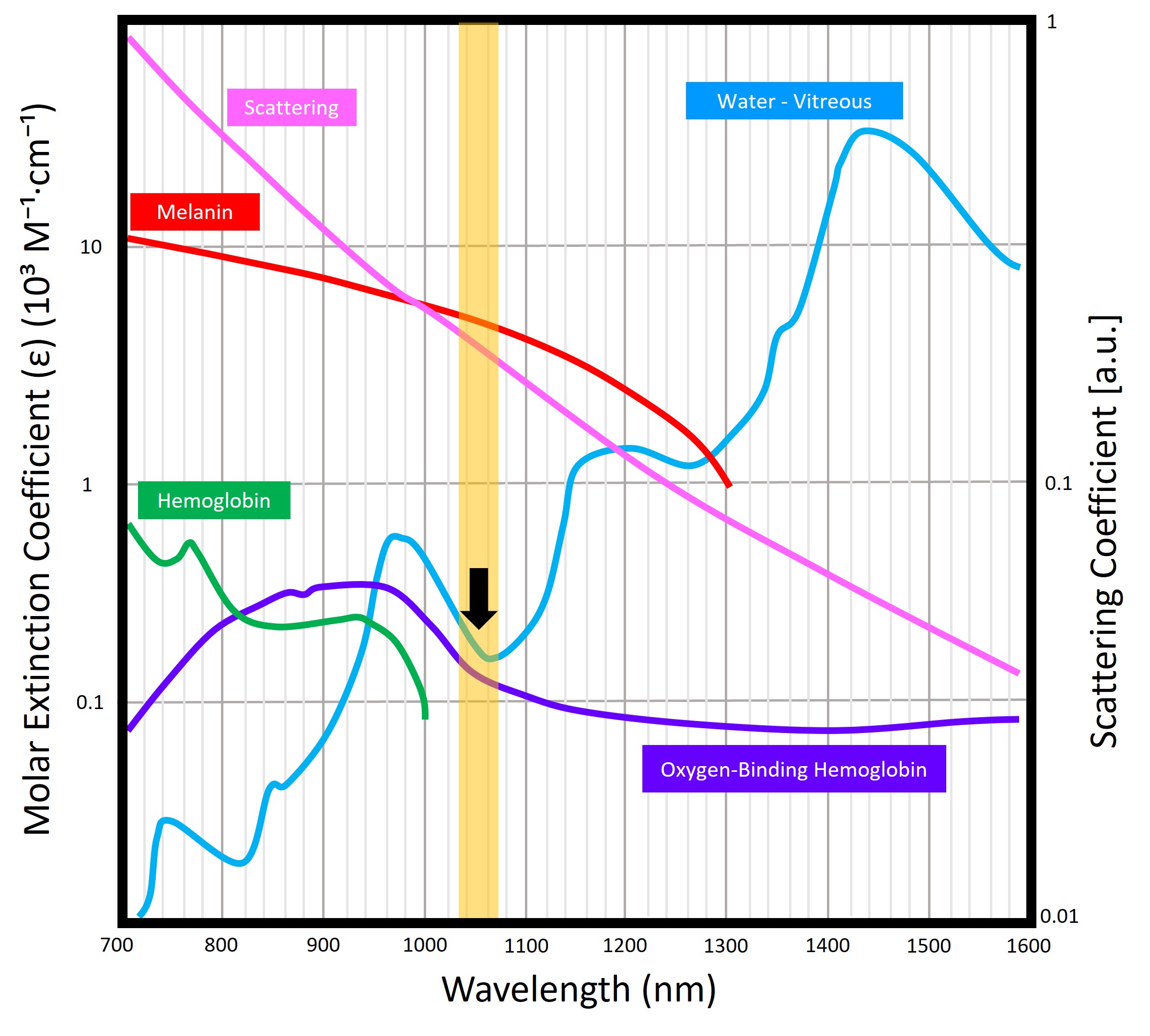
| Light Source | Uses a tunable laser around 1050 nm wavelength for deeper penetration and higher stability. | Uses a broadband super-luminescent diode (~840 nm), limited in depth and more affected by scattering. |
| Imaging Depth | Exceptional penetration through retinal pigment epithelium and into the choroid — visualizing deeper layers clearly. | Limited penetration; choroidal structures often indistinct or lost in shadow. |
| Scanning Speed | Much faster data acquisition, enabling quick, high-density scans with minimal motion artifacts. | Slower capture; longer scans often suffer from motion blur and patient discomfort. |
| Image Quality | Delivers consistent high-definition images across all tissue layers, even with cataracts. | Excellent for retinal surface imaging but signal quality drops with depth or opaque media. |
| Signal Stability | Laser source provides uniform signal strength throughout scans and across follow-ups. | Signal decays with depth; performance may vary over time. |
| Performance in Media Opacity | Penetrates through cataracts, corneal haze, or vitreous hemorrhage with minimal degradation. | More susceptible to scattering; image quality declines significantly in such conditions. |
| Measurement Reliability | Provides precise, repeatable quantitative data — ideal for long-term monitoring. | Reliable for superficial layers but less consistent for deep structures. |
| Technological Maturity | Represents the latest generation of OCT innovation; future-ready and expandable. | Older, well-established technology nearing its performance limits. |
| Maintenance & Calibration | Laser-based system offers better long-term stability and reduced recalibration. | Requires frequent calibration and periodic adjustments. |
| Clinical Advantage | Enables earlier disease detection, superior visualization, and greater diagnostic confidence. | Reliable for standard retinal scans but may miss subtle or deeper pathology. |
| Overall Value | Deeper • Faster • More Detailed • Future-Proof — the clear choice for advanced eye care. | Proven but technologically limited — less suitable for comprehensive imaging needs. |
Seven reasons it stands out in daily practice
VG200D is built for retina clinics and advanced diagnostics that demand high speed, deep penetration, widefield OCT/OCTA, and stable follow-up comparisons — in a workflow that stays fast and consistent.
High scan speed reduces motion artifacts and shortens exam time, improving patient comfort and throughput — ideal for high-volume days and repeat follow-ups.
Swept-source wavelength improves penetration and stability, helping clinicians visualize deeper layers more clearly — especially useful when media opacity is present.
Capture more retina per scan area to better assess peripheral involvement and reduce the need for multiple acquisitions, especially in complex cases.
Expanded OCTA montage supports better visualization of perfusion changes, non-perfusion areas, and vascular abnormalities across a wider retinal region.
Stable acquisition and consistent scan quality help clinics monitor progression and treatment response with confidence across repeated visits.
Integrating key imaging modes into one system streamlines workflow, reduces device switching, and supports comprehensive clinical documentation.
High-resolution structural OCT with widefield capabilities and strong repeatability makes VG200D a solid choice for advanced retina diagnostics, screening, and long-term disease monitoring — while keeping daily operation fast and scalable.