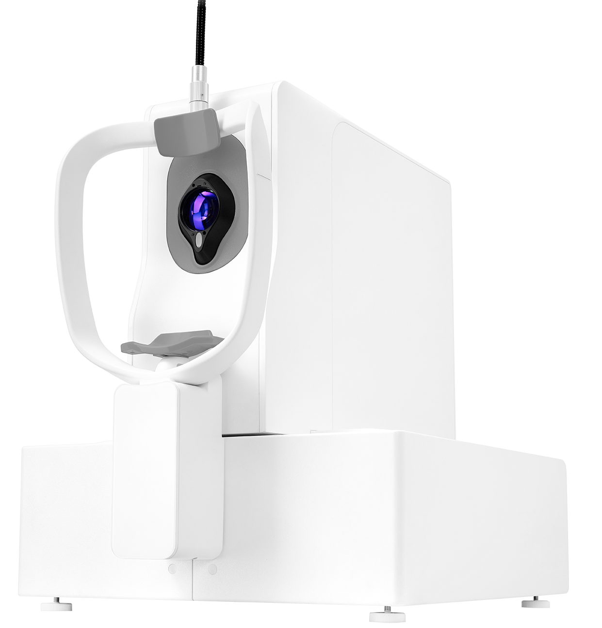
New OCT generation with anterior and posterior segment OCT and widefield posterior and anterior segment OCTA.
New OCT generation with anterior and posterior segment OCT and widefield posterior and anterior segment OCTA.

| DREAM OCT VG100D | ||
|---|---|---|
| OCT Imaging | ||
| Methodology | Swept-Source OCT | |
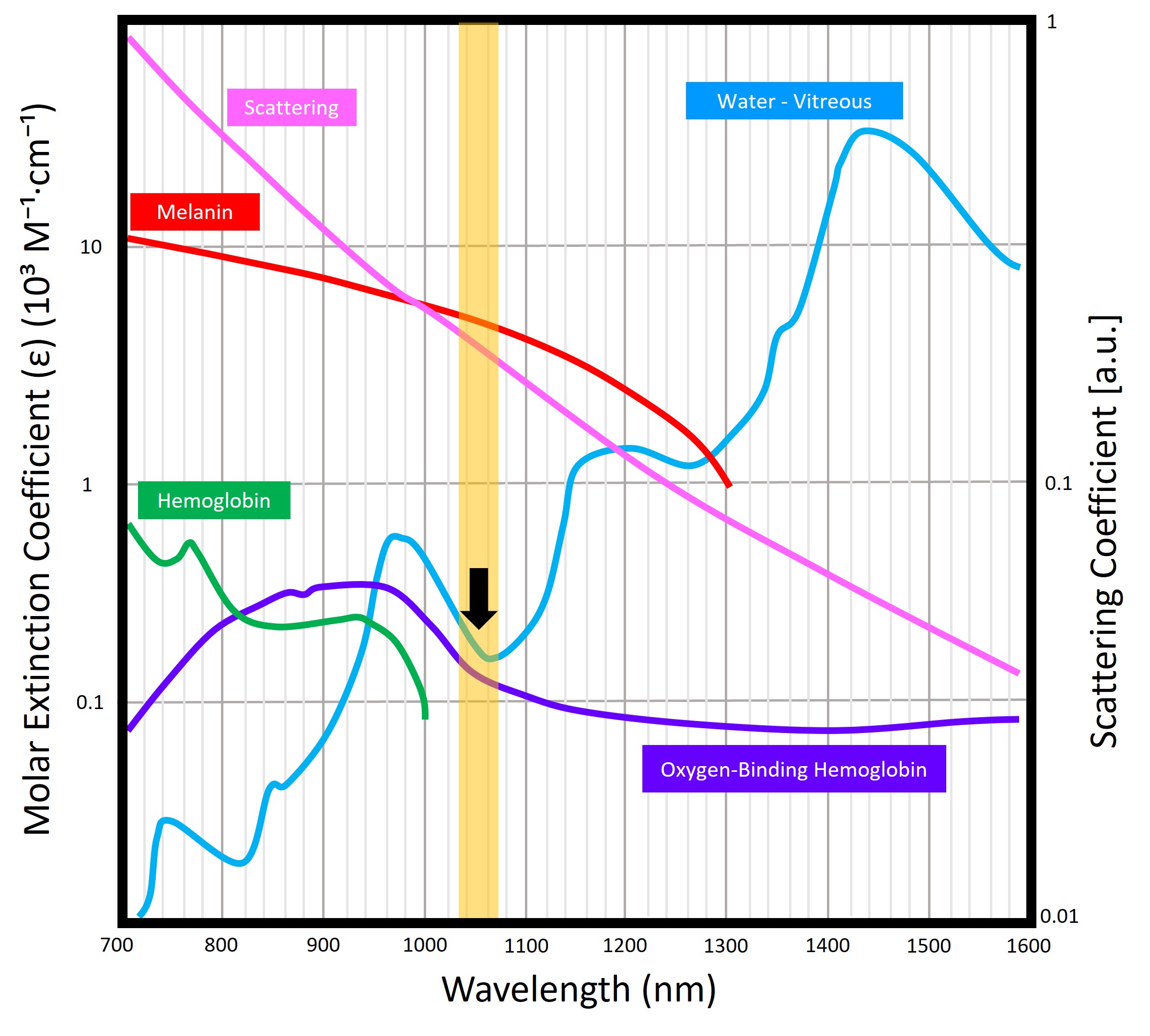
| OCT Central wavelength | 1030~1070 nm | |
| Scan speed | 100 kHz | |
| Axial resolution (Optical) | 5.5 μm | |
| Lateral resolution (Optical) | 15 μm | |
| A-scan depth | 9 mm (12.2 mm for AS) | |
| Scan range (Retina) | 83° (16 mm) | |
| Scan range (Anterior) | 20 mm | |
| OCTA Imaging | ||
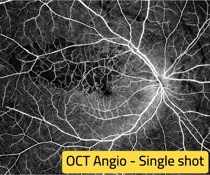
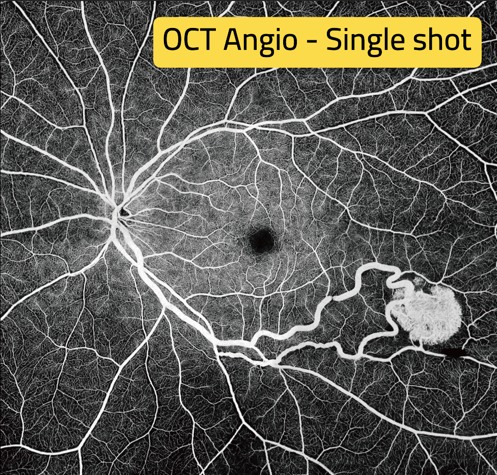
| Scan range (Retina) | 83° (16 mm × 16 mm) | |
| OCTA montage | 175° (34 mm × 30 mm) | |
| Algorithm | TRUE Angio™ | |
| Fundus Imaging | ||
| Methodology | cSLO | |
| Optical source | SLD | |
| Wavelength | 830±20 nm | |
| Field of view | 60° × 60° | |
| Others | ||
| Range of refractive compensation | -33 D ~ +40 D | |
| Alignment | Automatic / Electrical | |
| Feature | Swept-Source OCT | Spectral-Domain OCT |
|---|---|---|
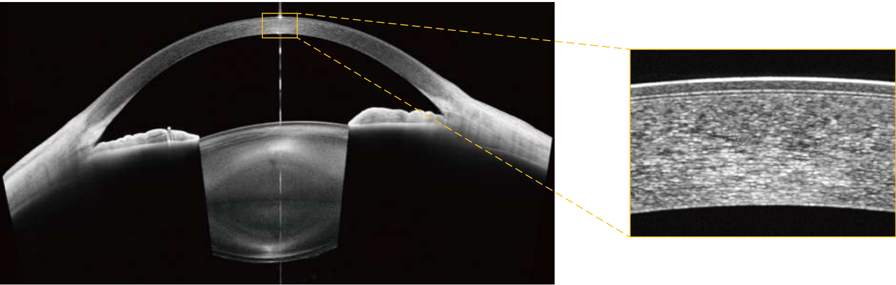
| Light Source | Uses a tunable laser around 1050 nm wavelength for deeper penetration and higher stability. | Uses a broadband super-luminescent diode (~840 nm), limited in depth and more affected by scattering. |
| Imaging Depth | Exceptional penetration through retinal pigment epithelium and into the choroid — visualizing deeper layers clearly. | Limited penetration; choroidal structures often indistinct or lost in shadow. |
| Scanning Speed | Fast data acquisition, enabling quick, high-density scans with reduced motion artifacts. | Slower capture; longer scans often suffer from motion blur and patient discomfort. |
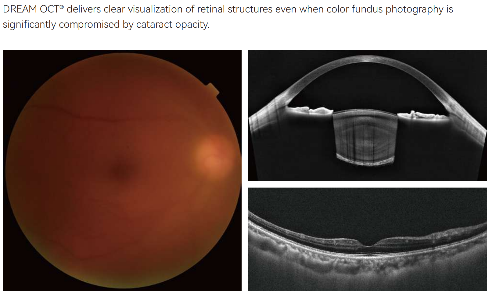
| Image Quality | Consistent high-definition images across tissue layers, even with cataracts. | Excellent for retinal surface imaging but signal quality drops with depth or opaque media. |
| Performance in Media Opacity | Better penetration through cataracts, corneal haze, or vitreous hemorrhage. | More susceptible to scattering; image quality declines significantly in such conditions. |
Seven reasons it stands out in daily diagnostics
VG100D is built for comprehensive, practical imaging — combining swept-source OCT/OCTA and fundus imaging with SuperDepth™ capability for deep anterior + posterior visualization, in an efficient 100 kHz workflow.
Balanced scan speed supports efficient patient flow with reduced motion artifacts — ideal for routine clinics, screening, and follow-up imaging.
Improved penetration and stable signal help clinicians evaluate deeper layers more clearly, especially when media clarity is not ideal.
Deep scan range supports broader structural assessment — helping visualize anatomy across depth for retina and anterior segment applications.
Expand vascular assessment beyond the posterior pole using montage imaging — supporting better evaluation of perfusion changes and peripheral involvement.
Use anterior and posterior segment OCT to support 360° angle assessment and comprehensive glaucoma evaluation, while enabling more precise optic disc cupping assessment (horizontal and vertical) than regular examination — helping clinicians document structures and monitor changes with confidence.
Combine key imaging modalities in one device to reduce switching, streamline documentation, and improve clinic efficiency.
VG100D supports anterior and posterior segment OCT, plus anterior and posterior segment OCTA, enabling comprehensive evaluation across the eye — including full assessment of the lens and cataract — in one efficient all-round platform.
Simple acquisition supports fast adoption by technicians and nurses in remote settings.
High-speed swept-source scanning improves workflow and reduces motion artifacts.
One platform covers anterior and posterior segment OCT for full eye evaluation.
Swept-source imaging improves penetration compared with spectral-domain, supporting difficult media.
Support glaucoma screening and follow-up with OCT/OCTA structural and vascular evaluation.
Assist lens/cataract assessment as part of a complete anterior segment workflow.
Identify structural and vascular pathology efficiently and document progression over visits. Assess the need for intervention without FFA.
Anterior segment OCTA supports detection and assessment of iris neovascularization.
Automated vault assessment supports safer phakic IOL follow-up in routine practice.
Full angle evaluation with a grading system helps guide management decisions.
Objective cupping assessment supports documentation and longitudinal glaucoma monitoring.
Overlay and detect PRP laser spots to assess ischemia and decide on augmentation needs.
Measure and compare FAZ between visits to monitor macular perfusion changes using OCTA.
Epithelial thickness mapping supports refractive evaluation and ocular surface assessment.
Lens tilt analysis supports advanced lens assessment and improved documentation.